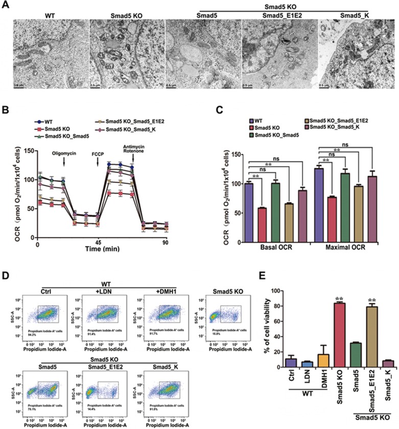
Figure 5.
Smad5 KO causes deficiency in mitochondrial respiration and malfunction of cellular bioenergetic homeostasis. (A) Electron microscopy images showing mitochondrial morphology in WT, Smad5 KO and pre-rescue hES. Scale bar, 0.5 μm. (B) Oxygen consumption rate (OCR) changes under mitochondrial stress in WT, Smad5 KO and pre-rescue hES as measured using the Seahorse Analyzer (n = 6). (C) Statistics of basal and maximal OCR in B. Data are represented as mean ± SEM of 6 independent experiments. Unpaired two-tailed Student's t-test. **P < 0.01. (D) Flow cytometry sorting of hES cells after propidium iodide staining. (E) Statistics of cell viability following serum withdrawal. Data are presented as mean ± SEM in three independent experiments. **P < 0.01.

