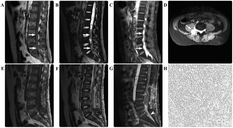
Figure 1.
In case one, the preoperative spinal magnetic resonance imaging scan revealed multiple masses (arrows) in the T12-S1 region. The masses were isointense on (A) T1-weighted and (B) T2-weighted images. (C) Following contrast agent administration, the masses demonstrated marked homogeneous enhancement. (D) The axial enhanced image revealed that the mass extended from the spinal canal into the paraspinal region. The postoperative (E) T1-weighted, (F) T2-weighted and (G) contrasted T1-weighted images demonstrated that the masses in the T12-L3 regions had undergone effective gross total resection, and that the mass (arrowhead) at the L5-S1 level had been partially resected for decompression. Pathological hematoxylin and eosin staining of the resected tissue revealed myeloid sarcoma (magnification, 200x). T12, thoracic 12; S, sacral; L, lumbar.

