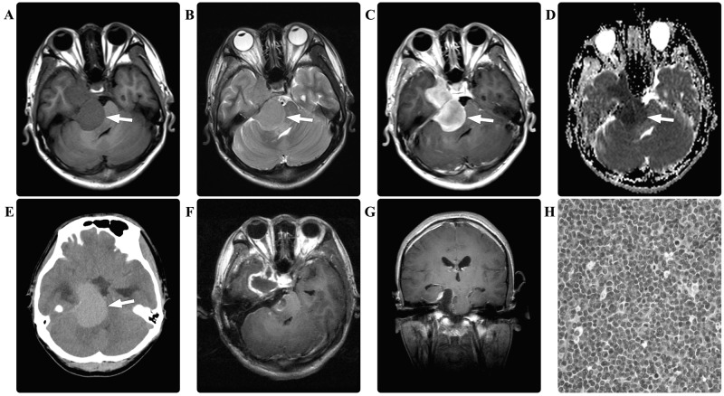
Figure 2.
In case two, the preoperative brain magnetic resonance imaging scan revealed a dumbbell shaped mass (arrows) in the right cerebellopontine angle, which appeared isointense on (A) T1-weighted and (B) T2-weighted images, with (C) marked homogeneous enhancement. (D) The diffusion-weighted imaging demonstrated restricted diffusion (arrow). (E) The computed tomography scan of the brain revealed a hyperdense lesion (arrow). Postoperatively, the (F) enhanced axial and (G) coronal T1-weighted images demonstrated a subtotal resection of the tumor. Pathological hematoxylin and eosin staining of the resected tissue revealed myeloid sarcoma (magnification, ×400).

