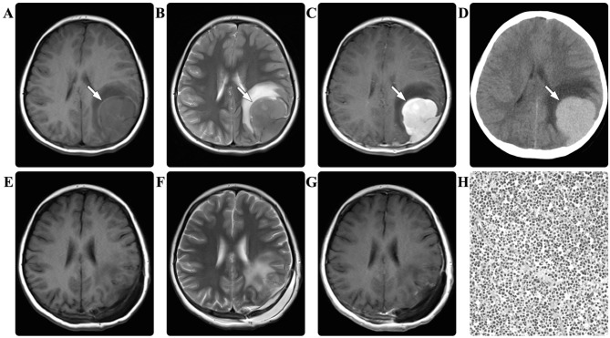
Figure 4.
In case four, the magnetic resonance imaging scan of the brain revealed a mass (arrows) in the left parietal lobe, which appeared isointense on the (A) T1-weighted and (B) T2-weighted images, with (C) marked homogeneous enhancement. (D) The computed tomography scan revealed that the mass (arrow) was hyperdense. The postoperative (E) T1-weighted, (F) T2-weighted and (G) contrasted T1-weighted images demonstrated that the mass had been subtotally resected. Pathological hematoxylin and eosin staining of the resected tissue revealed myeloid sarcoma (magnification, ×200).

