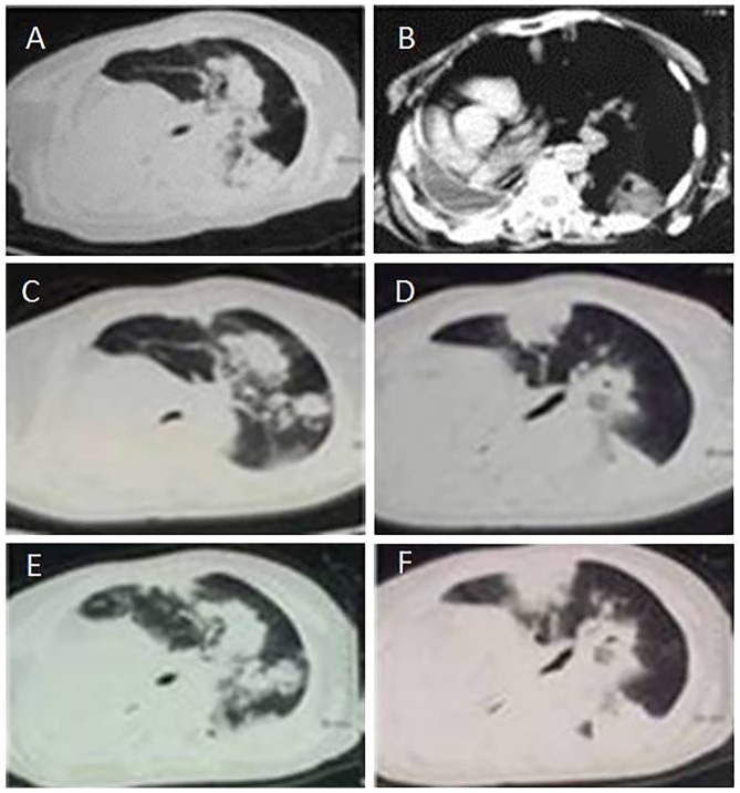
Figure 4.
CT scans of a patient with primary mucinous adenocarcinoma from 2015. (A) Thoracic CT scan following surgery on February 6, 2015, revealing a further increase in the number of metastases of the left lung; (B) Thoracic CT scan following surgery on February 16, 2015, revealing a pleural effusion of the right lung and a severe right deviation of the mediastinum; (C, D) CT scans following surgery on March 5, 2015, revealing a further increased number of metastases of the left lung; (E, F) CT scans following surgery on March 25, 2015, revealing a further increased number of metastases of the left lung. CT, computed tomography.

