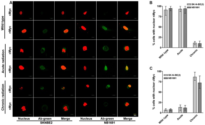
Figure 3.
Nuclear localization of cMYC in neuroblastoma cells treated with chronic radiation. (A) Localization of nMYC and cMYC proteins during radiation was identified using confocal fluorescent microscopy at ×10 magnification. Representative images illustrating nMYC and cMYC counterstained with propidium iodide. Under normal and acute radiation conditions, nMYC is observed to be localized in the nucleus, whereas cMYC is either absent or observed in low concentrations in the cytoplasm. Chronic radiation resulted in a significant increase in localization of cMYC in the nucleus, which is associated with a decrease in nMYC expression (scale bar, 10 µm). Cells were counted in five random fields and the average percentage of cells exhibiting nMYC and cMYC expression were determined. (B) There was a significant decrease in cells expressing nMYC in the nucleus and (C) an increase in cells expressing nuclear cMYC when treated with chronic radiation. Red staining indicates the nucleus; green staining indicates nMYC and cMYC. nMYC, v-Myc avian myelocytomatosis viral oncogene neuroblastoma derived homolog; cMYC, v-Myc avian myelocytomatosis viral oncogene homolog; Ab, antibody.

