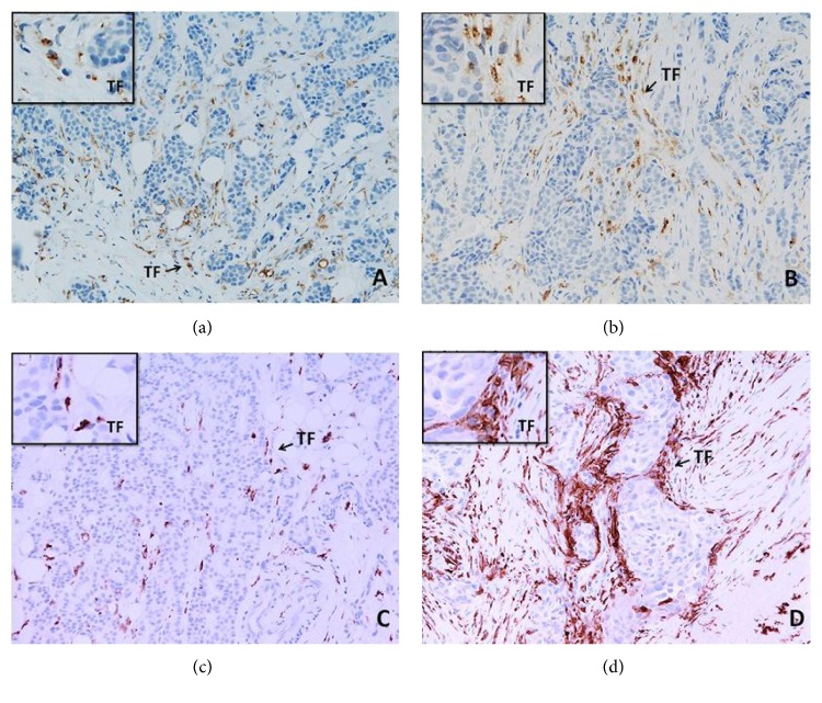
Figure 1.
CD68+ (a, b) and CD163+ (c, d) macrophages in the sections of LLABCs, using IHC staining, at 200x magnification. Briefly, heat-mediated antigen retrieval was performed using citrate buffer, pH 6 (20 mins). The sections were then incubated with MAbs to CD68 (Abcam, ab955) at a 1 : 300 dilution for 30 mins at RT, MAbs to CD163 (Abcam, ab74604) at a prediluted concentration for 30 mins at RT. Polymeric HRP-linker antibody conjugate was used as secondary antibody. DAB chromogen was used to visualize the staining. The sections were counterstained with haematoxylin. (a, c) Low level of CD68+ and CD163+ macrophage infiltration; (b, d) high level of CD68+ and CD163+macrophage infiltration. Tumours were classified as low level of infiltration when the positively brown membrane-stained cells were scattered or continuous along the tumour margin but did not extend from the tumour front (TF) for more than one cell layer. Extension for two or more layers from the TF was classified as a high level of infiltration.

