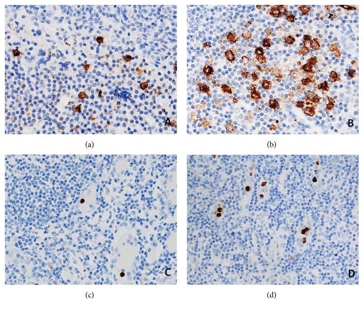
Figure 4.
CD1a+ DCs (a, b) and CD66b+ neutrophils (c, d) in the sections of axillary lymph nodes (ALNs), using IHC staining, at 400x magnification. Briefly, heat-mediated antigen retrieval was performed using citrate buffer, pH 6 (20 mins). The sections were then incubated with MAbs to CD1a (Dako, M3571) at a 1 : 200 dilution for 15 mins at RT, MAbs to CD66b (LSBio, LS-B7134) at a concentration of 10 μg/ml for 30 mins at RT. Polymeric HRP-linker antibody conjugate was used as secondary antibody. DAB chromogen was used to visualize the staining. The sections were counterstained with haematoxylin. (a, c) Low number of CD1a+ DCs and CD66b+ neutrophils; (b, d) high number of CD1a+ DCs and CD66b+ neutrophils. The average number of cell counts per HPF in tumour-free paracortical areas of ALNs with the greatest accumulation of the positive brown membrane-stained cells was quantified.

