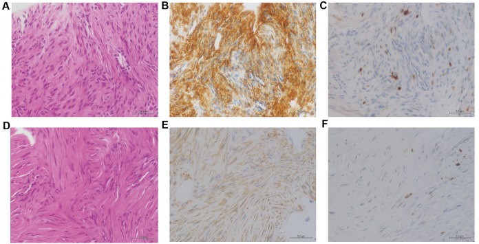
Figure 2.
Histological findings of the biopsy tissue specimen. (A-C) The histological findings of the biopsy specimen from case 1. (D-F) The histological findings of the biopsy specimen from case 2. (A and D) Results of H&E staining. The tissue samples exhibited a similar appearance consisting of interlaced bundles of spindle shaped tumor cells. (B and E) Immunohistochemical staining for α-SMA. The tumor cells each stained positive for α-SMA. (C and F) Immunohistochemical staining for Ki-67 antigen. Staining for Ki-67 revealed positive expression in <1% of the two tumors. Original magnification, ×400. H&E, hematoxylin and eosin. α-SMA α-smooth muscle actin.

