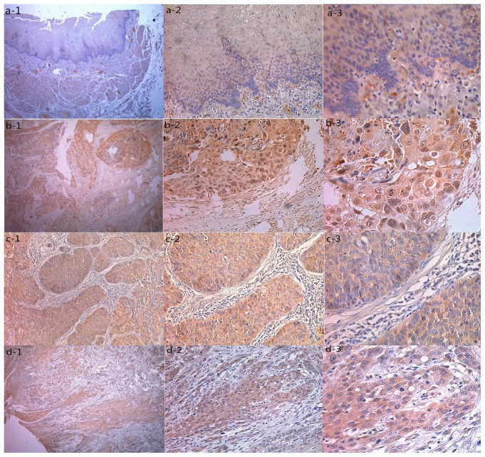
Figure 1.
Representative pattern of MAGE-A9 protein expression in ESCC and normal tissues in a tissue microarray. (A) Negative IHC staining of MAGE-A9 of non-cancerous tissue (magnifications: 1, ×40; 2, ×200; 3, ×400). (B) IHC staining of MAGE-A9 of well-differentiated ESCC tissue (magnifications: 1, ×40; 2, ×200; 3, ×400). (C) High IHC staining of MAGE-A9 of moderately-poorly differentiated ESCC tissue (magnifications: 1, ×40; 2, ×200; 3, ×400). (D) IHC staining of MAGE-A9 of well-differentiated ESCC sample (magnifications: 1, ×40; 2, ×200; 3, ×400). MAGE-A9, melanoma-associated antigen-9; ESCC, esophageal squamous cell carcinoma; IHC, immunohistochemistry.

