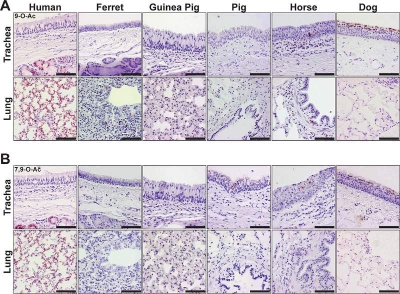
FIG 6 .

(A) Staining (red) of mammalian respiratory tissues for the distribution of the 9-O-Ac Sia, detected with the PToV-P4 HE-Fc SGRP. (B) Staining of respiratory tissues for the distribution of 7,9-O-Ac Sia, detected with the BCoV-Mebus HE-Fc SGRP. Both Sia forms were identified in respiratory tissues of all species examined, with variations in distribution and display. The 9-O-Ac and 7,9-O-Ac Sias generally appeared to have similar patterns of distribution. Human tissues displayed 7,9- and 9-O-Ac Sias in the tracheal submucosal glands and throughout the lung. These Sia forms were displayed at the tracheal epithelia of pigs, horses, and dogs. Bars, 50 μm.
