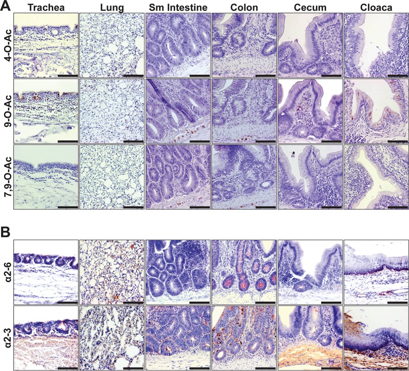
FIG 8 .

The distribution of Sias in the tissues of a Pekin duck, a major waterfowl reservoir of avian influenza viruses. (A) Staining (red) of respiratory tissues with SGRPs for O-acetyl modified Sias. Both 4-O-Ac and 9-O-Ac Sias are displayed at tracheal epithelia and in lung alveolar tissue. Within the digestive tissues, trace 7,9- and 9-O-Ac Sias are displayed in enteric ganglia of the intestines (Sm intestine, small intestine), whereas epithelial cells of the cloaca display abundant mono-9-O-Ac Sias. (B) Staining (red) of respiratory tissues for Sia linkage chemistry. Respiratory tissue displayed both Sia linkages, whereas digestive tissues showed more predominant α2-3-linked Sias. Bars, 50 μm.
