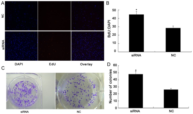
Figure 3.
PDCD4 knockdown promotes renal cancer cell proliferation. (A) EdU proliferation assay. Proliferating cells that have incorporated EdU are stained red, while the nuclei of all cells is stained blue with Hoechst 33342. All experiments were repeated three times. (B) Proliferative ability of 786-O cells following transfection with PDCD4 siRNA and NC siRNA. (C) Images of colony formation assay of 786-O cells transfected with NC siRNA and PDCD4 siRNA captured on day 14. (D) The colony-forming ability of 786-O cells was significantly increased in the PDCD4 siRNA-transfected group compared with the NC group 14 days after transcription. Student's t-test was used for comparisons of two independent groups. *P<0.05 vs. the NC group. PDCD4, programmed cell death 4; NC, negative control; siRNA, small interfering RNA.

