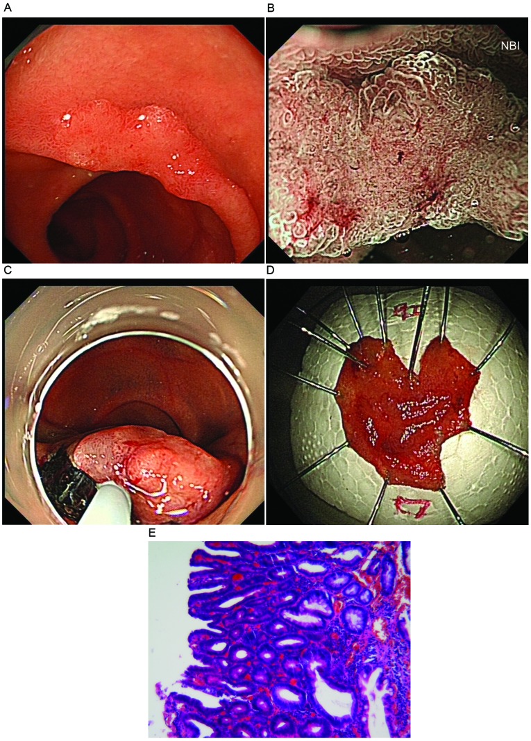
Figure 1.
An early superficial non-ampullary duodenal tumor. (A) A flat elevated lesion (1.2 cm) was visible in the second portion of the duodenum, using conventional endoscopy with white light imaging. (B) Surface pattern was preserved in one region and is absent in another region, and an unclassified vascular pattern was exhibited following magnifying endoscopy with narrow band imaging. (C) The endoscopic submucosal dissection method was performed to cure the lesion. (D) The whole lesion was cut from duodenum. (E) The lesion was diagnosed as a category 4/5 tumor by hematoxylin and eosin staining (magnification, ×100).

