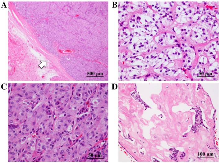
Figure 3.
Pathological findings of the tumor following hematoxylin and eosin staining. (A) The tumor was encapsulated by fibrous tissue (arrow). (B-D) The tumor was composed of cells with low-grade nuclear atypia, with (B) clear or (C) granular eosinophilic cytoplasm; a solid pattern of cell proliferation was observed, along with (D) hyalinized stroma.

