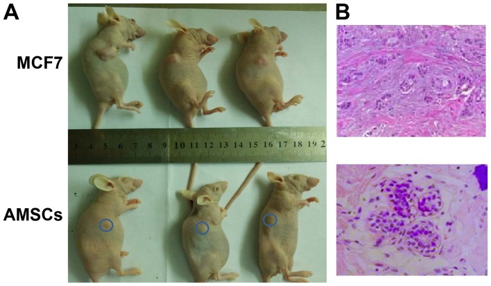
Figure 3.
To assess whether AMSCs are tumorigenic in vivo, AMSCs or MCF-7 breast cancer cells were subcutaneously injected into the dorsolateral area of nude non-obese diabetic/severe combined immunodeficiency-type mice (n=14 per group). (A) Representative mice from the AMSC group and the MCF-7 group were randomly selected at 30 and 14 days after injection, respectively, and photographed (blue circle: injection site in AMSC group). (B) Tissues from the injection site were isolated at 30 days (AMSC group) and 14 days (MCF-7 group) after injection and stained with hematoxylin and eosin. In the AMSC-injected tissue (magnification, ×400), normal mammary glandular structure was detected, in which the duct is lined with an inner layer of cuboidal luminal epithelial cells with an outer layer of squamous cells. MCF-7-injected breast tissue is shown on the right (magnification, ×200). AMSC, adult mammary stem cell.

