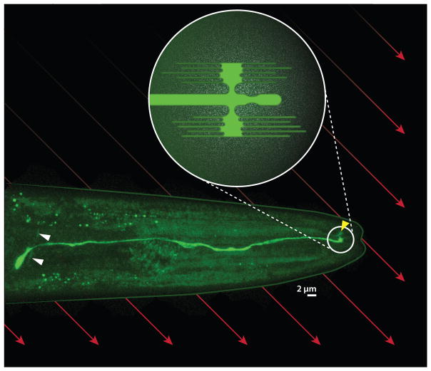
Figure 3.
AFD magnetosensory neuron in Caenorhabditis elegans. Fluorescent micrograph shows the right AFD neuron expressing a green fluorophore. The cell body has a curved axon (white arrowheads) that synapses onto other neurons and a dendrite that extends toward the tip of the worm’s head, where it features a complex sensory structure (circled). Reconstructions by White et al. (1986) and Doroquez et al. (2014) at the level of electron microscopy suggest that this structure includes a single cilium and antenna-shaped arrays of anterioposterior-directed microvilli on the dorsal and ventral sides (schematized in inset) Although most of the bilaterally symmetric left AFD neuron is out of view, its sensory structure is visible (yellow arrowhead). If the microvilli are associated with iron, the earth’s magnetic field (red arrows) may impose a mechanical force on the microvilli that depends on the orientation of the worm.

