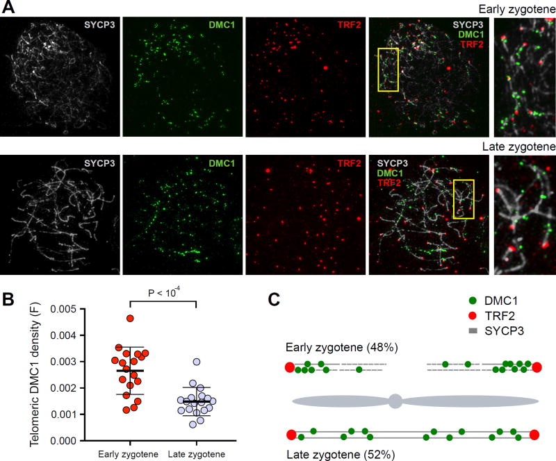
Figure 7. Increased frequency of DSB formation near telomeres.
(A) Early and late zygotene spermatocytes stained with SYCP3 (which detects axial elements) in grey, DMC1 (a marker of DSBs) in green, and TRF2 (a marker of telomeres) in red. DMC1 foci are clustered near telomeres at early zygotene. (B) Telomere-proximal DMC1 density is about 2-fold higher in early compared to late zygotene cells. For each cell, we manually traced all chromosome axes that unambiguously initiated at a TRF2 focus. We defined telomere-proximal regions as the 1μm of axis adjacent to the TRF2 focus (Fig. S30A). For each cell, the telomeric density of DMC1 foci (F) was calculated as ((Σ DMC1 foci in telomere proximal 1μm regions) / (total DMC1 foci) / (total TRF2 foci)). We do not count DMC1 foci adjacent to TRF2 foci that could not be unambiguously attributed to a particular chromosomal axis. This is particularly punitive for early cells as the DMC1 density appears highest in regions where TRF2 foci are clustered and individual axes are difficult to distinguish. Error bars indicate mean ± SD. P-values are calculated using a Mann-Whitney U-test. (C) Cartoon showing the distribution of DMC1 foci in early and late zygotene spermatocytes. 48% of zygotene cells are at the early stage where they show significantly higher frequency of DSB formation near telomeres. At late zygotene, DSBs are more evenly distributed. The combined signal from these two populations may result in the telomeric bias we observe in our genome-wide maps.

