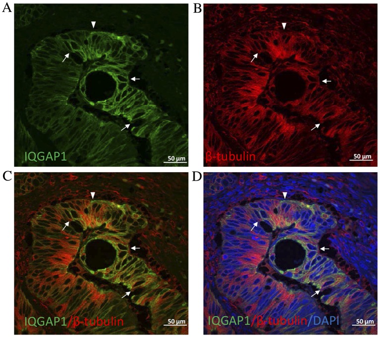
Figure 4.
Confocal microscopic analysis of (A) IQGAP1 (green) and (B) β-tubulin (red) expression by double immunofluorescence staining on CRC tissue sections. (C) IQGAP1 and β-tubulin colocalization and with (D) the addition of the nuclear stain DAPI. In the majority of cells, IQGAP1 co-located with microtubules at the cytoplasmic face of the nuclear envelope (arrows). Note that several cells exhibited increased IQGAP1 expression at the plasma membrane (arrowhead) at areas with no (or extremely low) tubulin expression. IQGAP1, IQ-motif containing GTPase activating protein 1.

