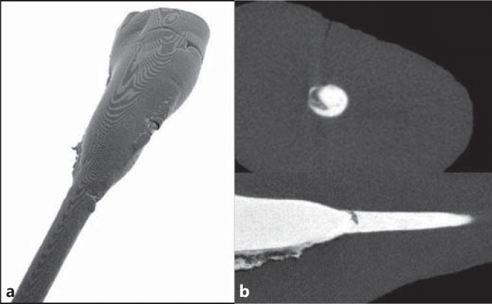
Fig. 1.
a Reconstructed 3D image of a tooth. b Perpendicular cross-sections of a root and an obturated root canal: background (black), dentine (dark grey) and filling material (white). Voids in the filling material are seen in 2D cross-sections as clusters of dark pixels surrounded by bright pixels.

