Fig. 1.

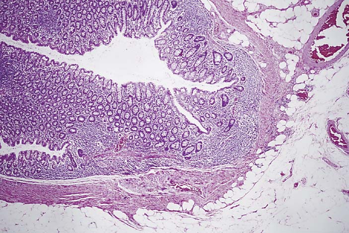
Low-power image of a right-sided diverticulum. HE. It shows a diverticulum with outpouching of the mucosa and submucosa with an attenuated layer of muscle. There is minimal associated chronic inflammation.

Low-power image of a right-sided diverticulum. HE. It shows a diverticulum with outpouching of the mucosa and submucosa with an attenuated layer of muscle. There is minimal associated chronic inflammation.