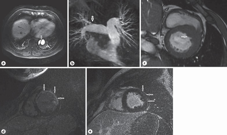
Fig. 1.
CMR images of the patients suspected of having ACS. a Contrast-enhanced T1-weighted images taken in an anatomical transversal orientation showing an intimal flap (arrow) in the thoracic aorta. b 3D-MR pulmonary angiography showing filling-defect signs (arrow) in the right upper lobe pulmonary arterial branch. c SSFP images in the short-axis view showing asymmetric increased thickness (arrow) at the base of the interventricular septum. d IR images in the short-axis view of the left ventricle showing transmural delayed contrast enhancement (arrows) in the extensive anterior wall. e IR images in the short-axis view of the left ventricle showing subepicardial delayed contrast enhancement (arrows) in the lateral wall.

