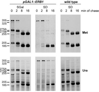
Figure 5.
Pulse–chase analysis of pre-rRNA processing. YMS0086 cells were grown in galactose-containing synthetic SGal medium (left) or transferred to glucose-containing SD medium for 20 h to deplete Erb1p (center). RNA from the control strain YMS0092 containing wild-type ERB1 is shown on the right. Cells were labeled with l-[methyl-3H]methionine (upper) or [5,6-3H]uracil (lower) for 2 min and chased with non-radioactive methionine or uracil, respectively, for 2, 8 and 16 min as described in Materials and Methods. The same total amount of RNA was loaded on each lane for methyl methionine-labeled samples and the same c.p.m. per lane were loaded for uracil-labeled samples. RNA was separated by formaldehyde–agarose gel electrophoresis, transferred to a nylon membrane and visualized by fluorography.

