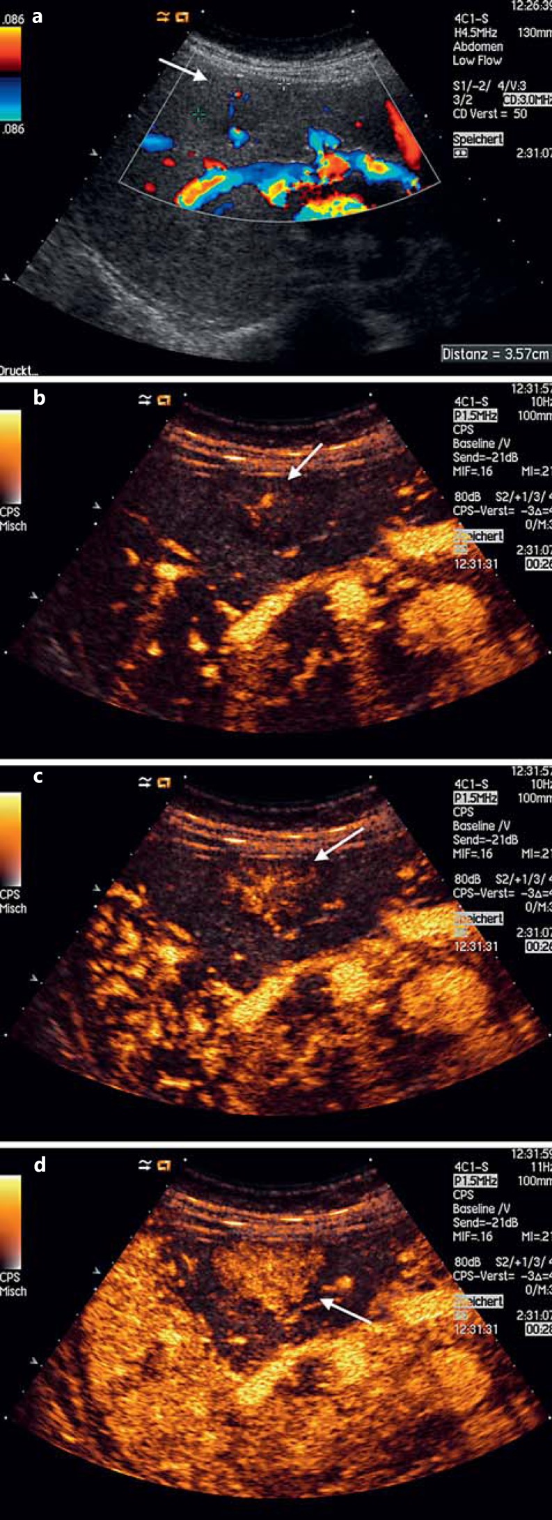
Fig. 2.
Focal nodular hyperplasia. a Discrete hypoechoic FLLs with central arterial vessel seen on color Doppler US. b, c ‘Spoke-wheel’ pattern of arterial centrifugal enhancement (26 s after intravenous CA administration), characteristic of a focal nodular hyperplasia. d Homogeneous enhancement (28 s after intravenous CA administration) rapidly seen after the first CA uptake within the lesion, also in favor of a focal nodular hyperplasia.

