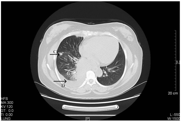
Figure 1.
CT scan of a 52-year-old female with stage IV primary pulmonary LELC. Arrow A, a CT scan revealed a large and well-defined soft tissue mass located in the right lower hilus of the lung (size, 6.5×5.0 cm). Arrow B, the bronchus in right lower lobe was narrowing with a thickened wall. Arrow C, multiple masses or nodules were observed in the right side of the chest wall, pleura and right oblique fissure with variable sizes (largest, ~1.4×2.7 cm). Arrow D, a limited amount of effusion was observed in the right side of the chest. CT, computed tomography; LELC, lymphoepithelioma-like carcinoma. HFS, head first-supine; MA, milliampere; KV, kilovolt; L, windows level; W, windows width; [L], length; GT, gradient; TI, time.

