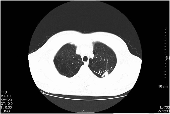
Figure 2.
CT scan of a 56-year-old male with stage IIIA primary pulmonary LELC. Arrow A, a CT scan identified a rough-edged, lobular and spiculated nodule in the apicoposterior segmental bronchus of the left upper lobe (size, 1.6×1.2 cm). Arrow B, pleural indentation was also observed. CT, computed tomography; LELC, lymphoepithelioma-like carcinoma. FFS, feet first-supine; MA, milliampere; KV, kilovolt; L, windows level; W, windows width; [L], length; GT, gradient; TI, time.

