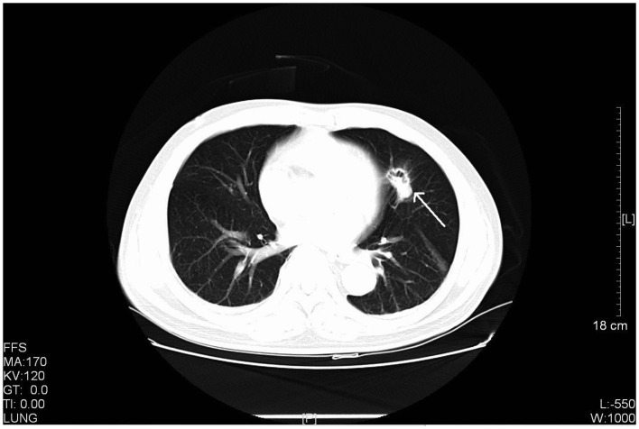
Figure 3.
CT scan of a 61-year-old male with stage IIIA primary pulmonary LELC. The CT scan revealed a rough-edged nodule in the lingular segment of the left upper lobe with cavity inside the lesion. CT, computed tomography; LELC, lymphoepithelioma-like carcinoma. FFS, feet first-supine; MA, milliampere; KV, kilovolt; L, windows level; W, windows width; [L], length; GT, gradient; TI, time.

