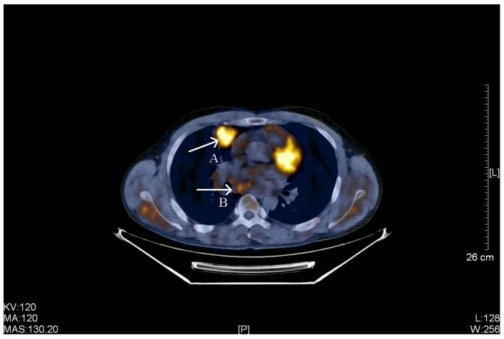
Figure 4.
PET-CT scan of a 38-year-old male with stage IIIB primary pulmonary LELC. Arrow A, a PET-CT scan identified a highly metabolic soft tissue (size, 3.5×3.4 cm) with slightly rough edge, lobular, spiculated, inhomogeneous density and pleural indentation in the anterior segment of the right upper lobe. Radioactive uptake increased and the SUVmax was 5.4. Arrow B, multiple enlarged lymph nodes in the right-side mediastinum and right hilus of the lung. The largest lymph node was located in the mediastinum (size, 2.4×2.2 cm), part integrated into clumps and radioactive uptake was slightly increased (SUVmax, 3.5). PET-CT, positron emission tomography-computed tomography; LELC, lymphoepithelioma-like carcinoma; SUVmax, maximum standardized uptake value. KV, kilovolt; MA, milliampere; MAS, milliampere sec; L, windows level; W, windows width; [L], length.

