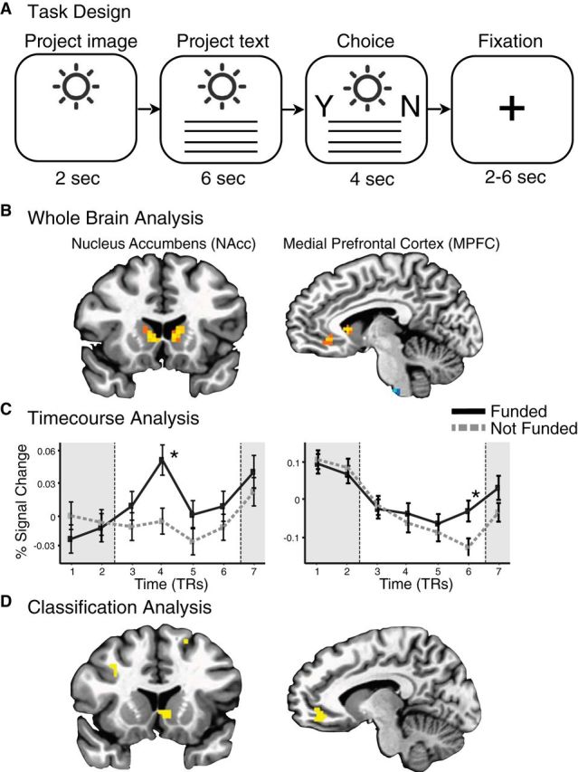
Figure 1.

Neural predictors of individual funding choices. A, Neuroimaging task trial design. Subjects saw a project image (2 s), project description (6 s), and spatially counterbalanced prompts to indicate their choice to fund or not (4 s), followed by a variable intertrial fixation interval (2–6 s). B, Whole-brain maps indicating neural activity associated with subjects' choices to fund projects. Warm-colored voxels are positively associated with choices to fund (vs not fund; p < 0.05, corrected). Significant clusters of voxels were observed in the bilateral striatum, including the NAcc), as well as in the MPFC. C, Time courses of neural activity extracted from bilateral NAcc (left) and MPFC (right) VOIs during the intertrial interval preceding each trial (TR 1–2; 4 s), project presentation (TR 3–6; 8 s), and choice period (TR 7; 2 s). Separate lines indicate trials in which subjects chose to fund (black, solid) versus not to fund (gray, dashed). Both regions show increased activity while viewing the project associated with subsequent choices to fund (*p < 0.05, corrected). D, Classification of individual funding choices. Whole-brain maps illustrate the top 1% of voxels that predicted individual choices to fund (yellow). As with whole-brain univariate analyses, this model-free classifier identified predictive voxel clusters in the NAcc and MPFC.
