Figure 1.
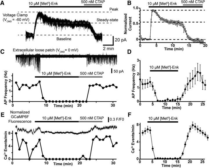
Sustained inhibition of AP firing and Ca2+ activity by MOR activation. A, Voltage-clamp recording from a POMC neuron showing attenuation of the GIRK current elicited by ME (10 μm) during a 10 min application. The residual GIRK current is eliminated the MOR selective antagonist CTAP (500 nm). Dotted lines are overlaid to highlight the baseline current (bottom dashed line), the peak GIRK current (top dashed line), and the steady-state GIRK current (middle dashed line) following desensitization. B, Summary data showing the attenuation of the GIRK current during prolonged ME exposure (n = 6 neurons, 4 animals). C, Extracellular loose-patch recording of a Pomc-Cre neuron expressing GCaMP6f during a 10 min ME (10 μm) application with subsequent reversal by CTAP (500 nm). Top trace, Action currents recorded in loose patch, with the bottom plot showing AP frequency over time. D, Summary data of the sustained inhibition of AP frequency by ME (10 μm; n = 6 neurons, 6 animals) over a 10 min application. E, Optical recording of the same neuron as in C. Top trace is the F/F0 normalized GCaMP6f fluorescence intensity with measured Ca2+ event frequency plotted below. In this cell, ME (10 μm) completely eliminated both AP firing and resolvable Ca2+ events. F, Summary data for the inhibition of Ca2+ events by ME (n = 29 neurons, 5 slices, 3 animals).

