
Figure 5.
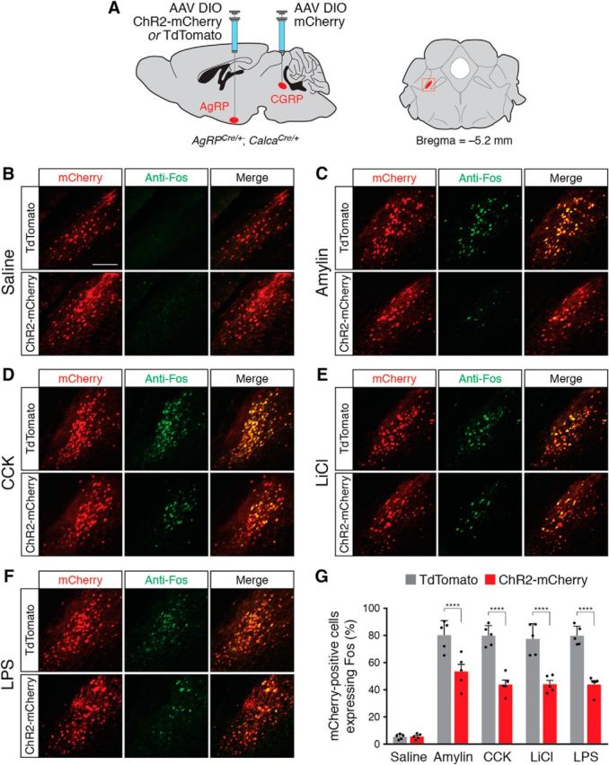
AgRP neuron stimulation reduces Fos expression in PBN CGRP neurons following administration of anorexigenic compounds. A, Injection strategy in AgRPCre/+; CalcaCre/+ double knock-in mice. Right, Coronal diagram representing the location of CGRP neurons; red square represents parabrachial region depicted in photomicrographs. B–F, Representative histological images showing coincident mCherry (red) and Fos (green) expression in CGRP neurons following injection of saline (B), amylin (C), CCK (D), LiCl (E), and LPS (F), in ChR2-mCherry- or TdTomato-transduced animals, following photostimulation of AgRP neurons. Scale bar, 250 μm. G, Quantification of coexpression of mCherry and Fos in the parabrachial nucleus. All values represent the mean ± SD, black dots represent individual animals (n = 5 animals per group). ****p < 0.0001, Bonferroni post hoc tests between TdTomato- and ChR2-mCherry-transduced animals.