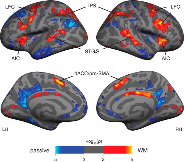
Figure 4.
Group-average activation (N = 15) for WM tasks (orange) versus sensorimotor control (blue). This analysis combines visual and auditory modalities. There is increased bilateral activation during WM in regions of lateral frontal cortex along the precentral sulcus and caudal inferior frontal sulcus, as well as in anterior insula, dorsal anterior cingulate/pre-SMA, STG/S, and the IPS and superior parietal lobule. The AIC and dACC/pre-SMA regions do not appear in the sensory modality contrast and are thus included in further analysis as putative “pure MD” regions.

