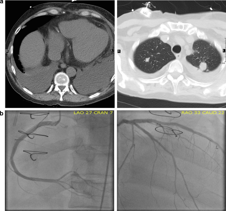
Fig. 1.
Cross sectional chest imaging. a CT scan of the chest showing bilateral pulmonary metastatic nodules, new onset of moderate bilateral pleural effusion but no evidence of significant pericardial effusion 5 days after administration of nivolumab. b Coronary angiography via left heart catheterization showing unchanged left main coronary artery, eccentric stenosis in the proximal LAD and patent stents in mid LAD and ramus intermedius. There was patency of the stents in the proximal-mid segments of LAD and ramus intermedius arteries but 70% stenosis in the distal right posterior descending artery consistent with findings on the most recent routine coronary angiography performed approximately 3 weeks prior to presentation

