
Fig. 1.
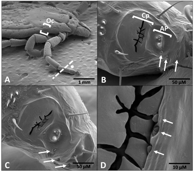
Scanning electron micrographs of Dermacentor variabilis Haller's organ (HO) and associated structures. (A) female, dorsal view at 25×, dotted line where tarsus I including HO was removed, (B) female, dorsal view of HO anterior pit and capsule at 500×, (C) male, dorsal view of HO anterior pit and capsule at 500×, and (D) female, dorsal view, aperture opening of capsule at 2500×. Arrows in panels B-D indicate undescribed structures resembling auricular or companiform sensilla that may serve as IR detectors or assist in this function in both male and female D. variabilis. The white star in panel A denotes the location of the HO (star just above structure). The ocellus (primitive eye) is located between the brackets in panel A. The dotted line denotes the location where the HO was ablated for the corresponding trials. Oc = ocellus, Cp = capsule, AP = anterior pit.