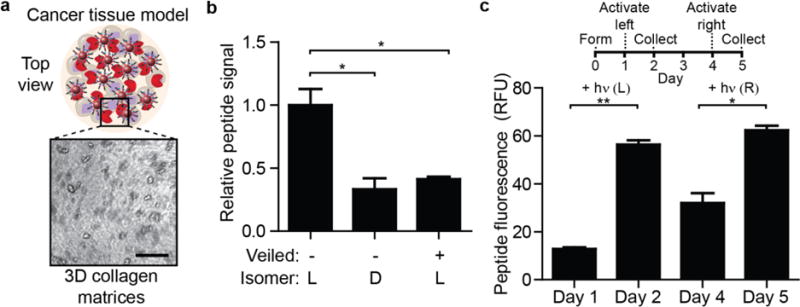
Figure 4. STREAMs embedded in cancer tissue models for protease sensing.

(a) 3D collagen tissues containing embedded colorectal cancer cells established as an in vitro model of the tumor microenvironment. Cells inside the collagen gel can be visualized and are homogeneously distributed (scale bar: 200 μm). C1-NPs (veiled or unmodified, L and D stereoisomers) were also embedded. (b) One day after forming the gel, the surrounding media was assessed for peptide fluorescence. Veiled substrates had significantly lower rates of proteolysis as did D-stereoisomer peptides compared to gels that contained L-stereoisomer particles (*P<0.05, two-tail Student’s t-test, n = 3, s.e.m.). (c) Spatial and temporal activation of STREAMs in cancer collagen tissue. The left half of gels was exposed to light on day 1 and total peptide signal was measured in collected supernatant. Three days later, the right half of gels was activated and peptide signal was measured (**P<0.01, *P<0.05, two-tail, paired Student’s t-test; n = 3, s.e.m.; light exposure: 30s at 200 mW/cm2).
