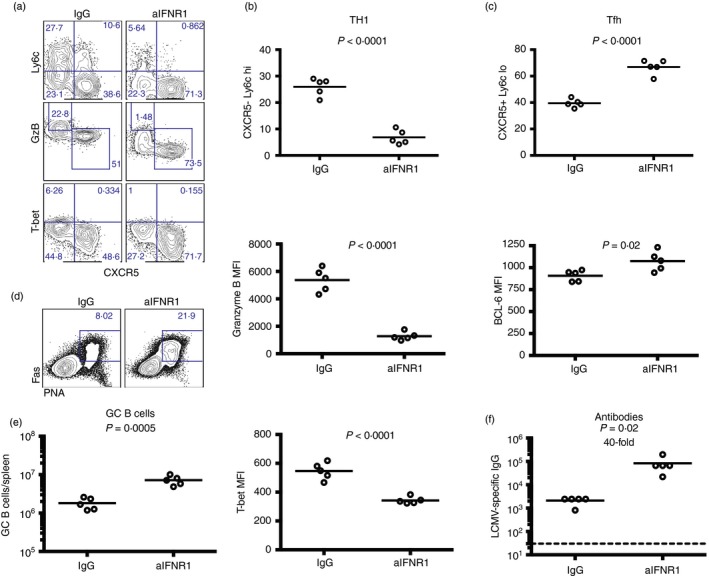
Figure 9.

Blockade of interferon type I signalling results in biased follicular T helper (Tfh) cell differentiation. (a) Representative FACS plots showing T helper subset differentiation following type I interferon blockade (gated on CD45·1+ SMARTA cells). (b) Percentage of SMARTA cells that are T helper type 1 (Th1) and their expression of granzyme B and T‐bet. (c) Percentage of SMARTA cells that are Tfh cells and their expression of Bcl‐6. (d) Representative FACS plots showing germinal centre (GC) B cells (gated from IgM− and IgD− B220+ CD3− live lymphocytes). (e) Numbers of GC B cells. (f) lymphocytic choriomeningitis virus (LCMV) ‐specific IgG responses by ELISA. 5 × 10⁶ naive SMARTA from spleen cells were transferred into congenically distinct recipient mice, followed by LCMV Cl‐13 challenge 1 day later, similar to Fig. 8. All panels represent data from spleen, except for (f), which is from sera. Data are from day 6 post‐infection. Data shown are from one experiment. Experiment was repeated with similar results, n = 5 mice/group per experiment. [Colour figure can be viewed at wileyonlinelibrary.com]
