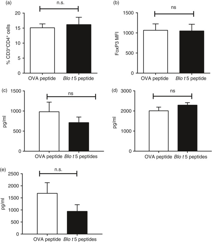
Figure 5.

Role of regulatory T cells and interleukin‐10 (IL‐10) in Blo t 5 peptide immunotherapy. (a) Infiltration of CD3+ CD4+ CD25+ FoxP3+ cells in lungs of mice that had received either Blo t 5 peptides immunotherapy or ovalbumin (OVA) peptide (mock) were assessed by flow cytometry (n = 14). (b) FoxP3 expression as measured by the MFI of FoxP3 staining in CD3+ CD4+ CD25+ FoxP3+ cells (n = 14). ELISA measurement of IL‐10 levels in (c) lymph node culture (n = 18) and (d) lung homogenate (n = 8) of mice that received peptide immunotherapy compared with control mice. (e) Levels of the T helper type 1 cytokine interferon‐γ as measured by ELISA (n = 18).
