
Figure 3. Identification of a Candidate OGT Intronic Splicing Silencer.
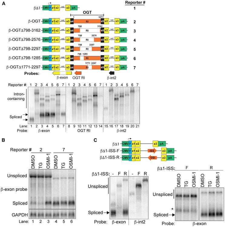
(A) Top: diagrams of reporters. Green, promoter and polyadenylation signals; blue, Flag-tag; yellow, β-globin exons; black line, β-globin intron 2; black, OGT exons; orange, OGT intron 4. Numbering is relative to the OGT intron sequence only; diagrams are not to scale. Positions of northern blot probes are also shown. Bottom: northern blot of RNA from cells transfected with the indicated reporter. The samples were loaded in parallel on the same gel. The membrane was subsequently cut (dashed lines) and hybridized to the indicated probe.
(B) Northern blot of RNA from 293A-TOA cells transfected with reporters and treated for 6 hr as indicated.
(C) Top: schematic of β-globin reporters with ISS in the forward (F) or reverse (R) orientation. Bottom: northern blots of reporter assays with transfected constructs, probes, and treatments as indicated. *Indicates an unknown transcript, likely a cryptic splice product. See also Figure S3.