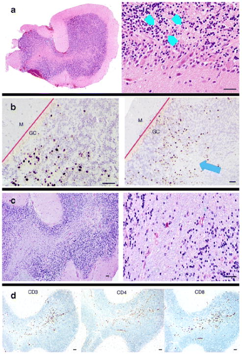
Fig. 3.
Pathology from open cerebellar biopsy. a, Hematoxylin and eosin staining shows a region of the granule cell layer with enlarged nuclei (blue arrowheads) next to normal-appearing granule cell neurons. b, Immunohistochemical staining of SV40 T antigen (ARUP Laboratories, Salt Lake City, Utah) was used to visualize JC virus-infected cells. Infected, positive staining cells are isolated to the granule cell (CG) layer. The molecular (M) layer is unaffected. The blue arrow highlights an area of focal granule cell loss. The red lines demarcate the boundary between the granule cell and molecular cell layers of the cerebellum. c, Luxol-fast blue/periodic acid Schiff (LFB-PAS) staining does not show demyelination, unlike PML. d, CD3, CD4, and CD8 immunohistochemical staining shows inflammatory T cell infiltrates affecting the granule cell layer. The pathology laboratory did not perform CD20 staining. The black scale bars at the bottom of each panel represent approximately 50 μM

