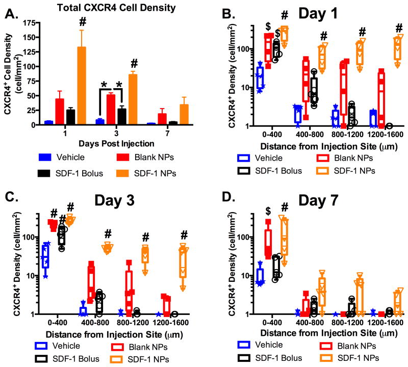
Figure 7.
Sustained release of SDF-1 incites transient dispersed CXCR4 activation. (A) CXCR4 cell density (cell/mm2) across the entire cortical ROI significantly increased with SDF-1 NPs compared to all groups at day 1 and 3 (#p < 0.01), then returned to control levels by day 7. At day 3, the blank NPs also elicited an increased CXCR4 cellular response compared to vehicle and bolus SDF-1 (A; *p < 0.05). (B-D) Spatial analysis demonstrated significantly increased and dispersed CXCR4 response with the SDF-1 NPs compared to all other groups at day 1 and 3 (B,C; #p < 0.01) and only at the most proximal region to the injection at day 7 (D; #p < 0.01). Both bolus SDF-1 and blank NPs elicited significantly increased CXCR4 within 400 μm of the injection compared to vehicle at day 1 (B; $p < 0.05) and compared to all other groups at day 3 (C; #p < 0.01). The blank NP cell density was significantly higher than vehicle at day 7 (D; $p < 0.05). Note: y-axis in B-D is log scale. Box and whisker plots used in B-D to demonstrate the span of data points within each group; n = 4-5 animals per group.

