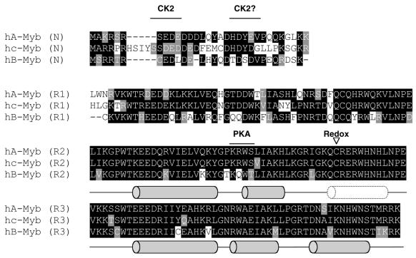
Figure 1.
The aligned amino acid sequences of the DBDs (NR123) of human A-, B- and c-Myb. Identical amino acids are marked with black boxes and similar amino acids are marked with grey boxes. The locations of the α-helices determined by NMR (28) are indicated as grey barrels and the flexible region (nascent helix) in R2 is indicated by a dotted barrel. The locations of kinase phosphorylation sites and redox-sensitive cysteines are indicated.

