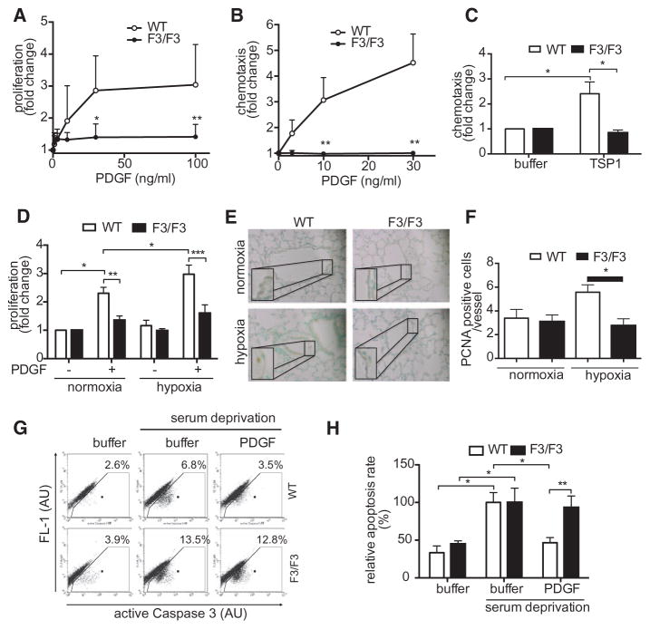
Figure 4.
Abolished platelet-derived growth factor (PDGF)–dependent proliferation and migration and enhanced susceptibility to apoptosis in F3/F3 pulmonary arterial smooth muscle cells (PASMCs). PDGF-BB–dependent proliferation (A) and PDGF-BB or thrombospondin (TSP-1)–induced migration (B and C) of PASMC from wild-type (WT) or β platelet-derived growth factor receptor (βPDGFR)F3/F3 mice were assessed by BrdU incorporation or with modified Boyden chamber assays, respectively. C, PDGF-dependent proliferation (BrdU incorporation) of PASMC was analyzed after exposure of cells to hypoxia (1% O2) or normoxia. D and E, Lung sections of WT and βPDGFRF3/F3 mice subjected to normoxia (21% O2) or hypoxia (10% O2) for 3 weeks stained for PCNA, counterstain with methylgreen. Representative experiment (D) and quantification of 3 independent experiments (E). F and G, Detection of apoptosis by flow cytometric staining of active caspase-3-positive cells. Representative experiment (F) and quantification of 3 independent experiments (G). AU indicates arbitrary units. *P<0.05, **P<0.01.

