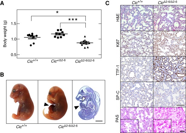
Figure 2.
Cic repressor activity is essential for mouse development. (A) Body weight of Cic+/+, Cic+/Δ2–6, and CicΔ2–6/Δ2–6 embryos at E18.5. Data represent mean ± SD. Asterisks depict statistically significant differences. (*) P < 0.05; (***) P < 0.001, unpaired Student's t-test. (B) Representative images of Cic+/+ (left) and CicΔ2–6/Δ2–6 (middle) embryos at E18.5. (Right) Hematoxilin and eosin (H&E) staining of a whole CicΔ2–6/Δ2–6 embryo section at E18.5. Bar, 2 mm. Arrowheads indicate omphaloceles. (C) H&E, Ki67, TTF-1, and SP-C immunohistochemistry (IHC) as well as periodic acid Schiff (PAS) staining in lung sections of Cic+/+ and CicΔ2–6/Δ2–6 embryos at E18.5. Bars: H&E, Ki67, TTF-1, and SP-C, 50 µm; PAS stainings, 25 µm. Inlays in PAS stainings represent magnifications.

