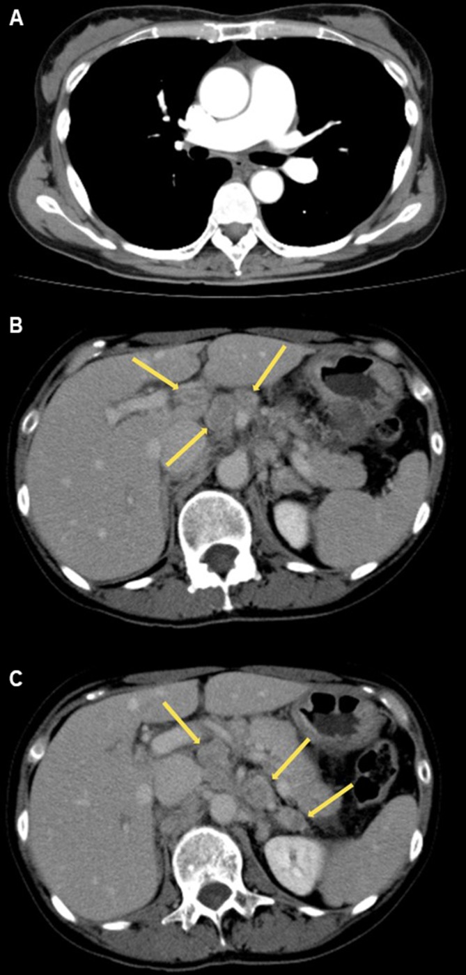
Figure 1.

CT on admission. (A) Mediastinal window image of lung field shows no thrombus in the pulmonary artery. (B) Lung window image shows no abnormality in the parenchymal parenchyma. (C,D) Mediastinal window image of abdomen shows enlargements of the para-aortic lymph nodes (yellow arrow).
