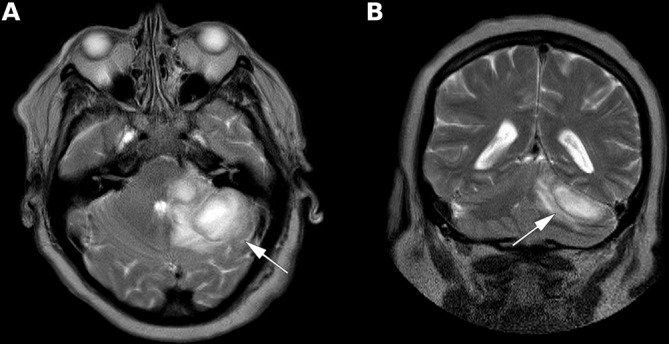
Figure 1.

Slides of the brain MRI show two cerebellar lesions (arrows): one in the left middle cerebellar peduncle (1.8 cm in size) and one in the left lateral hemisphere (3.5 cm in size). Both lesions were hypointense in T1 (not shown), hyperintense in T2 and were surrounded by vasogenic oedema. (A) Transversal plane. (B) Coronal plane.
