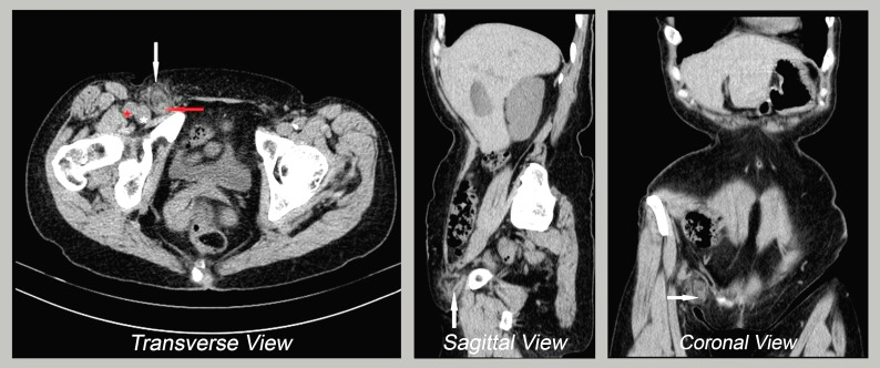
Figure 1.
Abdominopelvic CT scan. Transverse view: inflamed appendix (red arrow) inside the femoral ring (white arrow), medial to the femoral vein (white asterisk) and femoral artery (red asterisk). Sagittal view: tubular structure inside the hernia sac. Coronal view: inflamed appendix with adjacent fat stranding.

