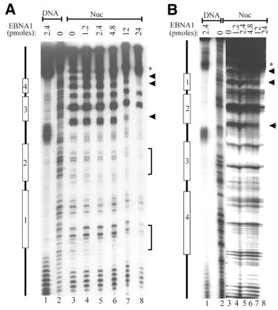
Figure 3.
DNase I footprints of ternary complexes. Nucleosomes containing DS DNA fragments (Nuc) were incubated with increasing amounts of EBNA452–641 to form ternary complexes, then subjected to DNase I footprint analysis. Bands protected by EBNA452–641 binding are indicated (arrows and brackets) as are DNase I-hypersensitive bands induced by EBNA452–641 (*). The DNase I digestion patterns of the naked DS DNA fragment (lane 2) and EBNA452–641 bound to the naked DS (lane 1) are also shown. The positions of EBNA1 binding sites 1–4 are indicated. (A) and (B) are footprints of opposite DNA strands.

