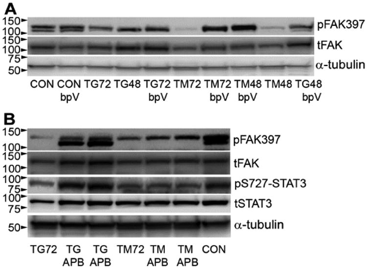
Figure 4. ER stress reduces pFAK through tyrosine phosphatases and calcium.

bEnd5 cells were treated with TG or TM for 48 or 72 h with or without the tyrosine phosphatase inhibitor bpV or calcium channel blocker APB.A) bpV prevented the loss of pFAK caused by ER stress, as shown in a Western blot. tFAK = total FAK, α-tubulin was used as a loading control. B) APB completely rescued pFAK against TG, but not against TM. This was accompanied by similar effects on pS727-STAT3. tSTAT3 = total STAT3. Blots in A and B are representative of n=3.
