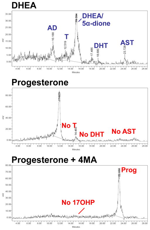
Figure 3.
Steroid flux from DHEA versus progesterone. LNCaP cells were treated with [3H]-DHEA (100 nM), [3H]-progesterone (100 nM), and [3H]-progesterone (100 nM) pretreated with 4-MA (10 μM), for 48 hours. Steroids were extracted from the medium, treated with glucuronidase and analyzed by thin-layer chromatography (not shown) or high-performance liquid chromatography (HPLC). DHEA is readily metabolized to androstenedione (AD), testosterone (T), dihydrotestosterone (DHT) and androsterone (AST). In contrast, for cells treated with progesterone (Prog), there was no detectable 17OH-progesterone (17OHP), T, DHT, or AST. No 17OHP is detected with pretreatment with the SRD5A inhibitor 4-MA, and little Prog metabolism occurs. These results suggest that the major metabolic pathway of Prog in LNCaP cells begins with 5α-reduction, rather than 17-hydroxylation.

