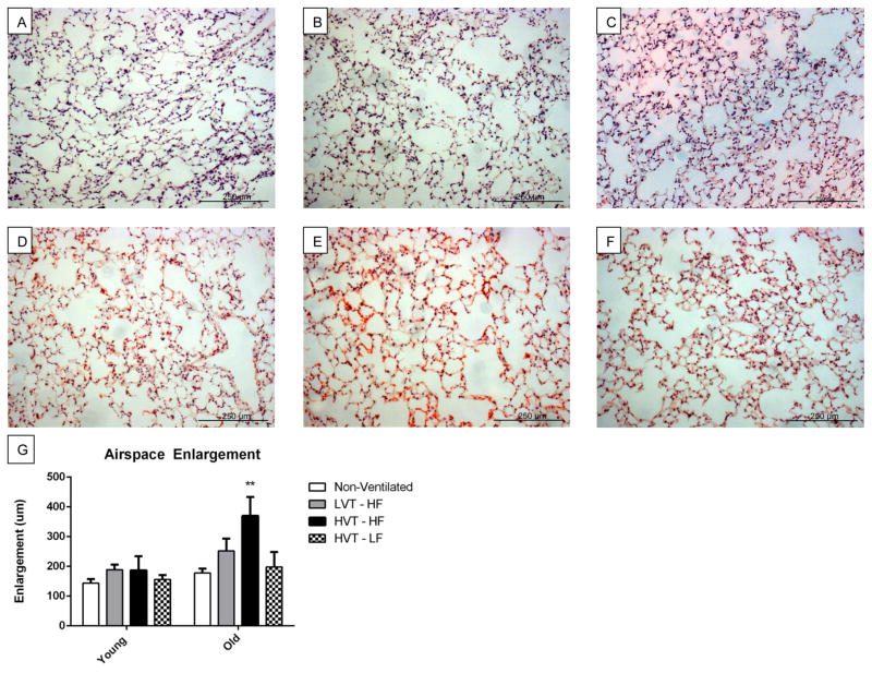
Figure 6.
A, B, C, D, E, F Representative 4Hr histological H&E images of A. Young LVT-HF, B. Young HVT-HF, C. Young HVT-LF, D. Old LVT-HF, E. Old HVT-HF, and F. Old HVT-LF lung sections respectively. G. Airspace Enlargement. There was a statistically significant groupwise difference across age with old groups having significantly greater enlargement than young. Enlargement was greater in the Old HVT-HF group than in all others. Data are presented as mean +/− st.dev N = 3 for Young Non-Vent, 6 for Young LVT – HF, 6 for Young HVT – HF, 3 for Young HVT – LF, 3 for Old Non-Vent, 3 for Old LVT – HF, 4 for Old HVT – HF, 3 for Old HVT – LF **p<0.01.

